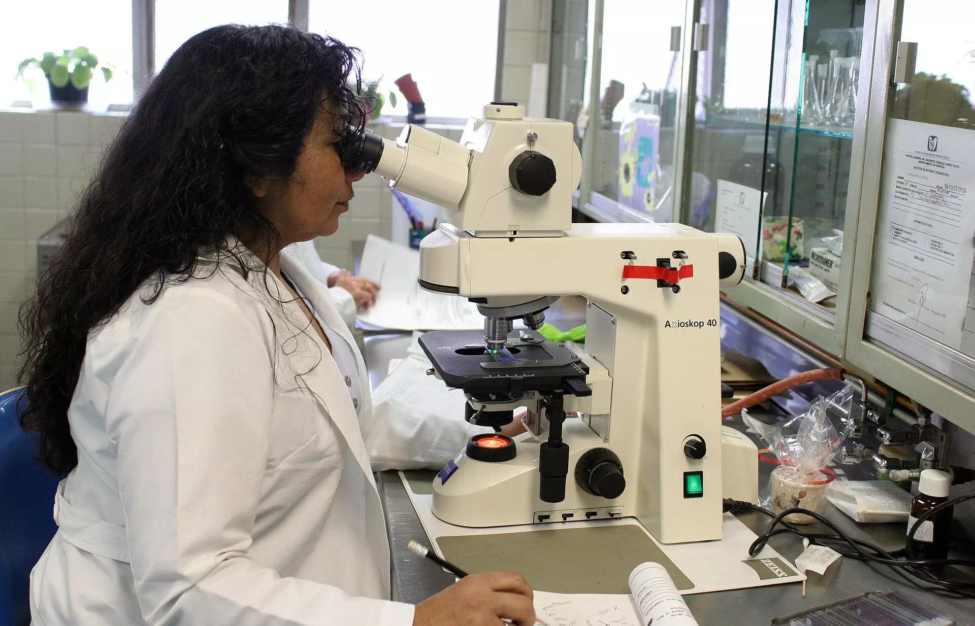
When observing biological samples under a microscope, it is crucial to consider the medium in which the lens of the objective is situated. The disturbance in the light beam occurs when the lens is in a different medium than the sample being viewed. For instance, if the lens is surrounded by air while viewing a watery sample, the light rays bend more sharply in the air than in the water. This discrepancy in refractive indices leads to a misleading measurement of the depth in the sample, causing it to appear flattened.
Over the years, various theories have been developed to address this issue of depth distortion in microscopy. However, early theories assumed a constant corrective factor for determining depth, regardless of the distance of the sample from the lens. It wasn’t until the 90s that Nobel laureate Stefan Hell highlighted the possibility of a depth-dependent scaling factor. Associate Professor Jacob Hoogenboom from Delft University of Technology emphasizes the importance of this revelation in understanding the true depth of biological samples.
Former postdoc Sergey Loginov, in collaboration with Delft University of Technology, has conducted groundbreaking research to prove the depth-dependency of the corrective factor. Through meticulous calculations and a mathematical model, Loginov demonstrated that the sample appears more flattened closer to the lens than farther away. This significant discovery has been further validated by Ph.D. candidate Daan Boltje and postdoc Ernest van der Wee in laboratory experiments.
The implications of this research extend beyond theoretical understanding, as it has practical applications in the field of microscopy. By accurately determining the depth of biological samples, researchers can precisely isolate proteins and their surroundings for detailed analysis with electron microscopy. This advancement significantly reduces the time and cost involved in microscopy experiments, allowing for more efficient exploration of relevant proteins and biological structures.
Researcher Daan Boltje emphasizes the importance of accurate depth determination in studying proteins within biological systems. Understanding the precise structure of proteins is essential for unraveling abnormalities and diseases, paving the way for more effective treatment and prevention strategies. The depth-dependent scaling factor plays a crucial role in ensuring that researchers are examining the right biological targets with precision.
To facilitate the practical implementation of depth-dependent scaling factor, the research team has developed a web tool and software for researchers to calculate the corrective factor specific to their experiments. By inputting relevant details such as refractive indices, aperture angle of the objective, and light wavelength, researchers can determine the depth-dependent scaling factor for their microscopy studies. This tool also allows for the comparison of results with existing theories, providing a comprehensive analysis of depth distortion in microscopy.
The depth-dependent scaling factor in microscopy represents a significant advancement in the field of biological research. By accurately determining the depth of samples, researchers can enhance the precision and efficiency of their experiments, ultimately leading to a deeper understanding of biological structures and mechanisms. The collaboration between theoretical insights and practical tools has opened up new possibilities for studying proteins and combating diseases with greater accuracy and effectiveness.


Leave a Reply