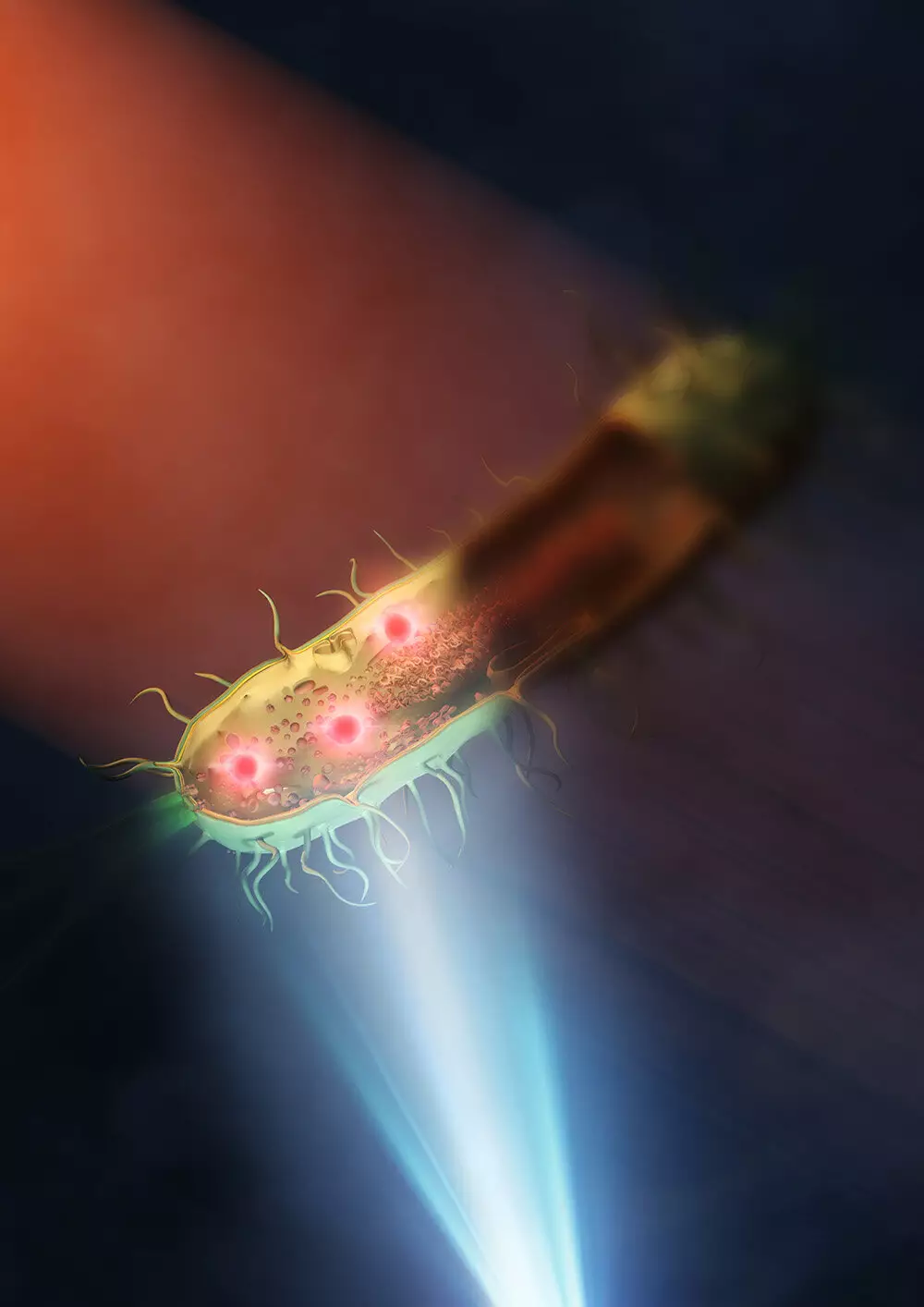
In a groundbreaking development at the University of Tokyo, a team of researchers have successfully enhanced the capabilities of mid-infrared microscopy, allowing for unprecedented insights into the structures within living bacteria at the nanometer scale. Published in Nature Photonics, this advancement marks a significant improvement in resolution, with images now produced at a remarkable 120 nanometers, representing a 30-fold enhancement compared to traditional mid-infrared microscopes.
Despite the marvels of modern microscopes that enable us to explore the microscopic world of viruses, proteins, and molecules, each imaging technique comes with its own set of limitations. For instance, super-resolution fluorescent microscopes necessitate the labeling of specimens with fluorescence, which can be harmful to samples and may lead to sample bleaching with prolonged exposure to light. Electron microscopes, while capable of providing intricate details, require samples to be placed in a vacuum, making it impossible to study live samples. On the other hand, mid-infrared microscopy offers the unique advantage of providing both chemical and structural information about live cells without the need for staining or damaging them, making it an appealing tool for biological research.
With its historical constraints in resolution, mid-infrared microscopy has often taken a backseat in biological research due to its limited capabilities compared to other imaging technologies. While super-resolution fluorescent microscopy can achieve resolutions down to tens of nanometers, mid-infrared microscopy has typically been limited to around 3 microns. However, the team of researchers at the University of Tokyo overcame this hurdle by achieving an exceptional spatial resolution of 120 nanometers, a significant milestone that significantly surpasses previous limits. Professor Takuro Ideguchi from the Institute for Photon Science and Technology at the University of Tokyo highlighted the breakthrough, emphasizing that the team’s synthetic aperture technique involving multiple images taken from various illuminated angles played a crucial role in enhancing the resolution.
To address the challenge of light absorption by traditional lenses used in mid-infrared microscopy, the researchers innovatively placed the samples, which included bacteria such as E. coli and Rhodococcus jostii RHA1, on a silicon plate that reflected visible light and transmitted infrared light. By eliminating the need for two lenses and utilizing a single lens setup, the team significantly improved the illumination of the sample with mid-infrared light, resulting in clearer and more detailed images of the intracellular structures of bacteria. Professor Ideguchi expressed astonishment at the level of detail achieved, envisioning the potential applications of the enhanced spatial resolution in studying critical issues such as antimicrobial resistance.
Looking ahead, the researchers are optimistic about further advancements in mid-infrared microscopy, with the potential to refine the technique in various aspects. By leveraging superior lenses and shorter wavelengths of visible light, the spatial resolution could potentially be further refined to below 100 nanometers, opening up new possibilities for high-resolution imaging in biological research. The ability to visualize samples at such a detailed scale holds tremendous promise for investigating a wide range of topics, from infectious diseases to cellular mechanisms, paving the way for more accurate and comprehensive scientific discoveries in the future.
The recent breakthrough in mid-infrared microscopy represents a significant leap forward in biological research, offering unparalleled capabilities to observe living cells with remarkable clarity and precision. With ongoing advancements and improvements in this innovative imaging technique, the future of microscopy looks brighter than ever, promising new insights and discoveries that may revolutionize our understanding of the microscopic world.


Leave a Reply