Exposure to mercury (Hg) is notoriously known for its extreme neurotoxic effects, even to those who study mercury compounds themselves. Renowned physicist Michael Faraday experienced firsthand the detrimental consequences of prolonged exposure to mercury vapors, which forced him to halt his research due to deteriorating health. Another tragic example is lab chemist Karen Wetterhahn, who lost her life to dimethylmercury poisoning after a few drops came into contact with her latex-gloved hand. These incidents serve as a somber reminder of the inherent risks associated with mercury exposure.
While numerous studies have focused on the exposure and effects of mercury in marine and sea creatures, the question arises: can mercury ions find their way into the brains of terrestrial animals? Initially skeptical, Dr. Yulia Pushkar, a professor of Physics and Astronomy at Purdue University’s College of Science, sought to explore this intriguing query. With her brain imaging program established since 2008 and a skilled research group specializing in sample preparation, measurements, and data analysis, Pushkar’s team collaborates with researchers worldwide. Recently, their groundbreaking findings were published in Environmental Chemistry Letters, revealing the presence of mercury in the brains of mongooses collected in Okinawa Island.
Unraveling the Mystery of Mercury in Mongoose Brains
While the discovery of mercury in mongoose brains raises many concerns, the exact source of this toxic element remains unclear. Several possibilities exist, including contaminated water, consumption of bird eggs, mineral exposure, or even airborne mercury particles. However, one indisputable fact emerges from this research: the presence of mercury in the brains of terrestrial animals is an alarming indication. As Dr. Pushkar explains, “Hg is very toxic at low concentrations as Hg can bind and affect the function of essential biomolecules. Efficiency of detoxification will depend on uptake and binding constant inside detected accumulations and potential leakage from these if brain cells die. As of now, there is no known way to safely dissolve these aggregates from tissue, and there are no reports of reversing Hg poisoning of the neural system. The main approach we should all take is to avoid any exposures, especially chronic ones like in Faraday’s case.”
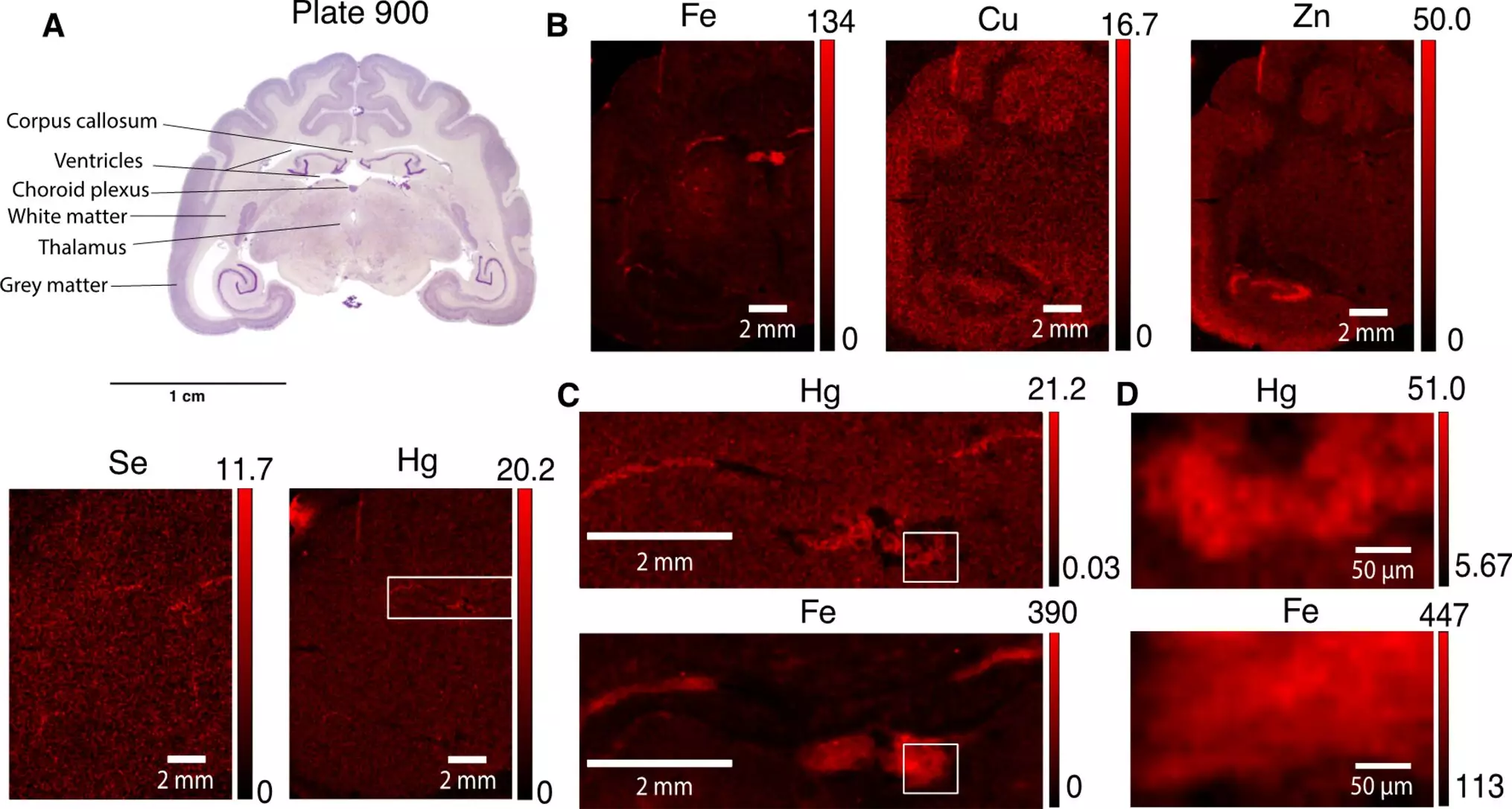
Dr. Pushkar initially held doubts regarding the detectability of mercury in the brains of mongooses. Neurotoxic elements typically exist in ultra-low concentrations, even if they manage to infiltrate the brain. To overcome her skepticism, the research team turned to the Advanced Photon Source at Argonne National Laboratory, where the mongoose brains underwent intense X-ray exposure. Contrary to expectations, the scans demonstrated the presence of mercury within the brain samples.
After meticulous scanning and analysis of the brain samples, Pushkar’s team identified specific brain cells that exhibited higher mercury concentrations. Cells of the choroid plexus, responsible for forming the blood-cerebrospinal fluid barrier, and astrocytes of the subventricular zone contained punctate structures rich in mercury. These structures measured between 0.5 and 2 microns in size. Further research is needed to uncover which selenium-containing biological molecules bond with mercury within these cells. The research team comprised Pavani Devabathini and Gabriel Bury, both graduate students, and Darrell Fischer, who was an undergraduate student at the time (now attending Harvard graduate school). The data collection and analysis were collaborative efforts by the entire team.
Implications for Environmental Monitoring and Human Health
This remarkable discovery holds significant implications for environmental monitoring of terrestrial animals and provides new tools for tracing mercury in brain cells. It has long been established that human activities result in the emission of 2,000 metric tons of mercury compounds annually, and yet the fate of this neurotoxic element remains uncertain. While most previous studies focused on the impact of mercury on marine biota, such as fish and whales, this research highlights the adverse effects on terrestrial species. Dr. Pushkar notes that it is likely the human brain reacts to mercury in a similar fashion through interactions with the cells of the choroid plexus and astrocytes.
The presence of mercury in the brains of terrestrial animals raises significant concerns regarding the potential health risks for both wildlife and humans. The findings of Dr. Pushkar and her team shed light on an often-overlooked aspect of mercury exposure and emphasize the importance of minimizing our contact with this toxic element. Further research is necessary to fully understand the mechanisms of mercury accumulation in brain cells and to develop effective methods for detoxification.
Leave a Reply