Scientists from the University of Tokyo led a team of researchers who have created a new method of imaging and analyzing biological samples. The new imaging method, called RESORT, combines the benefits of two leading technologies: super-resolution fluorescence imaging and vibrational imaging. While super-resolution fluorescence imaging provides good spatial resolution, vibrational imaging compromises spatial resolution but can use a broad range of colours to help label many different constituents in cells. The new imaging technique uses something known as Raman scattering, a special interaction between molecules and light which helps identify what’s in a sample under the microscope. The team successfully performed RESORT imaging of mitochondria in cells to validate the technique. The study was published in the journal Science Advances.
Details of the new imaging technique
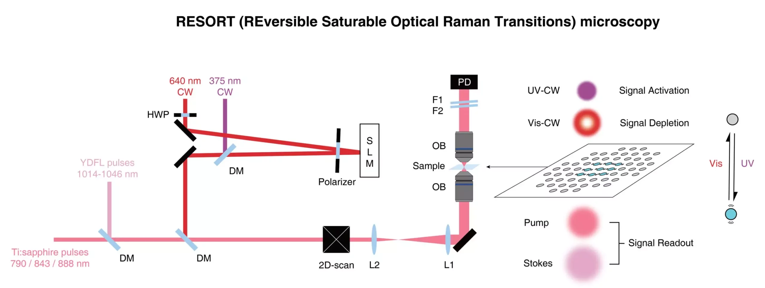
The new RESORT imaging technique is a laser-based technique that uses reversible saturable optical Raman transitions. The technique involves several stages, and while it might seem complicated, the setup is less complicated than that of the techniques it’s aiming to replace. Firstly, the specific components of the sample to be imaged need to be labeled with special chemicals called photoswitchable Raman probes, whose Raman scattering can be controlled by the different kinds of laser light employed by RESORT. Next, the sample is placed within an optical apparatus used to correctly illuminate the sample and build an image of it. The sample is then irradiated with two-color infrared laser pulses for detecting Raman scattering, ultraviolet light and a special donut-shaped beam of visible light. Together, these constrain the area where Raman scattering can occur, which means the final stage, imaging, can detect the probe at the very precise point, which leads to a high spatial resolution.
Impact of the new imaging technique
The team’s main aim was to improve microscopic imaging for use in medical research and related areas. The new RESORT imaging technique will be able to image many components in living samples in action to analyze complex interactions like never before. This will contribute to a deeper understanding of fundamental biological processes, disease mechanisms and potential therapeutic interventions. It is not just about gaining higher-resolution images of microscopic samples; after all, electron microscopes can image these things in far greater detail. However, electron microscopes necessarily damage or impede the samples they observe. Through the future development adding more colours to the palette of Raman probes, RESORT will be able to image many components in living samples in action to analyze complex interactions like never before. The advancements made in the design of the laser could be used in other laser applications as well, where high power or precise control is required, such as materials science.
The team of scientists has created a new imaging technique, RESORT, that combines the benefits of two leading imaging techniques, super-resolution fluorescence imaging and vibrational imaging. The technique involves several stages, including labeling the specific components of the sample to be imaged with photoswitchable Raman probes and irradiating the sample with various laser pulses. The new imaging technique will contribute to a deeper understanding of fundamental biological processes, disease mechanisms and potential therapeutic interventions. Furthermore, the advancements made in the design of the laser could be used in other laser applications as well, such as materials science.
Leave a Reply