In a recent study published in Nature Communications, researchers from Swansea University have developed a new imaging method for neutral atomic beam microscopes. This innovative technique has the potential to revolutionize the way engineers and scientists scan samples, leading to faster results and improved image resolution.
The existing neutral atomic beam microscopes used a pinhole scanning method, where the sample is illuminated through a microscopic pinhole and the scattered beam is recorded to build an image. However, this approach has a major limitation – the imaging time required, as the image is measured one pixel at a time. Additionally, reducing the pin-hole dimension to improve resolution results in a dramatic decrease in beam flux and longer measurement times.
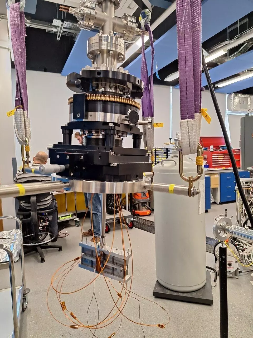
The research group led by Professor Gil Alexandrowicz from the chemistry department at Swansea University has developed a new and faster alternative method to pinhole scanning. They demonstrated the new method using a beam of helium-3 atoms, a rare light isotope of regular helium. This method involves passing a beam of atoms through a non-uniform magnetic field and using nuclear spin precession to encode the position of the beam particles interacting with the sample.
Ph.D. student Morgan Lowe built the magnetic encoding device and conducted the initial experiments, which confirmed the efficacy of the new method. The team used numerical simulations to show that the magnetic encoding method could improve image resolution with a smaller increase in time compared to the traditional pin-hole microscopy approach.
Professor Alexandrowicz highlighted the potential of the new method to enhance image resolution without significantly increasing measurement times. He also mentioned the possibility of new contrast mechanisms based on the magnetic properties of the sample. The team plans to further develop the method to create a fully operational prototype magnetic encoding neutral beam microscope for testing resolution limits, contrast mechanisms, and operation modes.
The development of the new imaging method for neutral atomic beam microscopes by Swansea University researchers represents a significant advancement in the field of microscopy. This innovative technique has the potential to streamline the imaging process, improve resolution, and enable new contrast mechanisms. The future looks promising for neutral beam microscopy with the introduction of this groundbreaking method.


Leave a Reply