In the intricate world of cellular biology, ribosomes play a pivotal role as the site for protein synthesis, a fundamental process that drives numerous biological functions. Recent advancements by researchers at the University of Tsukuba shine a light on the complexities of this process through innovative modeling techniques. The ribosome synthesizes proteins by translating messenger RNA (mRNA) into polypeptide chains, which later fold into functional proteins. Understanding the environment within ribosomes is crucial for revealing how proteins achieve their three-dimensional structures, which are essential to their functionality.
Historically, our grasp of protein synthesis has been limited by insufficient models that inadequately represent the ribosome’s internal environment. Previous studies identified that proteins may begin to adopt critical 3D structural features while still navigating through the tubular channel known as the ribosome tunnel. However, the mechanisms governing this process remained elusive. Recognizing the need for a more refined approach, the researchers at the University of Tsukuba conducted a thorough analysis of the various ribosomal tunnel structures documented in scientific literature.
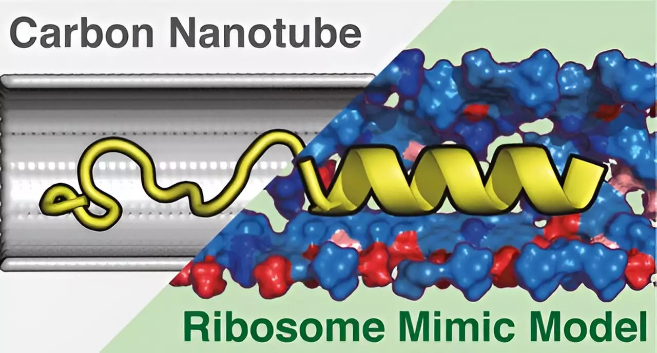
The result of their analysis prompted the creation of the Ribosome Environment Mimicking Model (REMM), a cylindrical model designed to replicate not only the inner dimensions but also the vital chemical characteristics of ribosome tunnels. This innovative model marks a significant step forward, allowing for more nuanced simulations of how proteins interact with the ribosome’s internal environment. To offer a comparative basis, the researchers also constructed a conventional carbon nanotube (CNT) model, which, while able to replicate the physical dimensions of the tunnel, falls short of including the chemical diversity found in actual ribosomes.
Utilizing molecular dynamics simulations, the team rigorously examined how proteins behave within these differing models. Their findings indicated that the REMM significantly surpassed the CNT model in generating protein structures that closely resembled those observed experimentally in biological settings. The chemical diversity of the REMM was pinpointed as a crucial factor in its enhanced accuracy. This highlights the importance of not just physical dimensions, but also the biochemical environment in which protein synthesis occurs.
This research not only enhances our understanding of ribosomal function but also paves the way for more advanced studies into protein conformations within living cells. As the REMM continues to be refined, it holds the potential to unravel deeper insights into the processes of translation and protein folding. The implications of such advancements are vast, potentially informing the development of more effective therapeutic strategies for diseases linked to protein misfolding and malfunction.
The contributions from the University of Tsukuba stand as a testament to the power of computational modeling within biological research. By closely mimicking the ribosome’s internal environment, these developments stand to revolutionize our understanding of protein synthesis processes. The journey from simplistic models to highly detailed simulations signifies a bright future for the field of molecular biology, where our comprehension of life’s building blocks continues to evolve.


Leave a Reply